A Clinician’s Guide to Urology Treatment: Symptoms, Diagnostics, and Procedural Care
Introduction
Stepping into a urology clinic often comes after days or months of dealing with uncomfortable, ambiguous symptoms. From a clinician’s viewpoint, the urinary and male reproductive systems are among the most responsive yet sensitive networks in human physiology. When disruptions arise—whether sharp flank pain, progressive urinary hesitancy, or subtle changes identified during routine screening—the primary clinical goal is straightforward: clarify the root pathology swiftly and build a targeted care strategy. Modern medical pathways allow practitioners to move far beyond generalized interventions. Today’s urology treatment encompasses a wide spectrum of interventions, ranging from structured active observation and pharmacological therapies to outpatient endoscopic procedures and robotic surgery. Navigating this landscape requires understanding how specialists analyze diagnostic data, how clinical facilities are organized, and how individual health profiles directly shape procedural decisions.
Understanding Urological Conditions and Healthcare Pathways
The genitourinary tract coordinates two vital roles: renal clearance, which maintains biochemical and fluid balance, and male reproductive physiology. When these systems falter, clinical presentation varies significantly based on anatomical depth.
Practitioners generally categorize urological care into subspecialized divisions: endourology (managing intraluminal blockages such as calculi), urologic oncology (addressing malignancies of the kidney, bladder, prostate, and testes), functional and female urology (treating voiding dysfunction, prolapse, and incontinence), and andrology. Patients seeking clinical evaluation often span every age group. A young adult may present with sudden infection or stone colic, while older patients frequently require evaluation for parenchymal prostate expansion or urothelial alterations. Clinical evaluation ensures benign conditions are managed conservatively while suspicious lesions receive timely, systematic staging.
Common Symptoms and Warning Signs: A Diagnostic Perspective
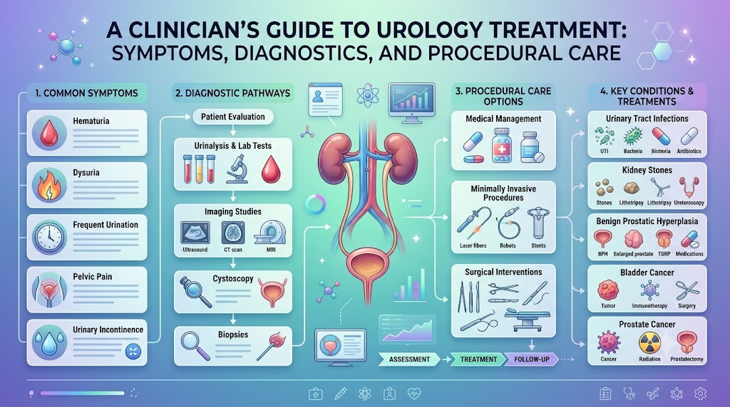
In urology, symptoms rarely indicate a single diagnosis. Instead, they serve as clinical signals pointing to potential functional or structural issues:
- Irritative Lower Urinary Tract Symptoms (LUTS): Sudden urgency, increased urinary frequency, and nocturia often indicate localized detrusor muscle irritation, mucosal inflammation, or incomplete bladder emptying.
- Obstructive Voiding Signs: Straining, urinary hesitancy, a weak or intermittent stream, and terminal dribbling typically suggest mechanical resistance at the bladder outlet or within the urethra.
- Dysuria: Discomfort or burning sensations during voiding usually indicate bacterial infection, chemical irritation, or urethral inflammation.
- Hematuria: Blood in the urine—whether macroscopically visible as red or cola-colored, or identified microscopically on lab dipsticks—warrants prompt, thorough investigation to exclude malignant or structural origins.
- Flank and Pelvic Discomfort: Acute, colicky costovertebral angle pain reflects ureteral distension from stone obstruction, whereas deep suprapubic or perineal dullness often points to prostate or bladder inflammation.
A complete clinical history clarifies the timeline, severity, and functional impact of these symptoms, guiding the selection of targeted diagnostic tests.
Causes and Contributing Risk Factors
Evaluating urological pathology involves identifying how systemic and local factors interact:
- Age-Associated Structural Remodeling: As systemic endocrine profiles change over time, prostate stromal and epithelial cells naturally proliferate, and bladder compliance gradually shifts.
- Genetic and Familial Tendencies: Inherited metabolic variations can predispose individuals to hypercalciuria and nephrolithiasis, while specific genetic markers increase vulnerability to urological malignancies.
- Metabolic and Cardiovascular Status: Vascular disease, hyperlipidemia, and diabetes alter microvascular perfusion to the pelvic organs, contributing to neurogenic voiding changes and erectile dysfunction.
- Prior Trauma and Instrumentation: Historical catheterizations, pelvic surgeries, or severe past infections can cause fibrosis, stricture formation, and drainage impairments.
These clinical variables help physicians determine whether a condition is localized and transient or part of a systemic, chronic process.
Diagnostic Protocols and Medical Tests
Specialists avoid standardized testing packages in favor of a stepwise diagnostic process, with each assessment answering a specific clinical question:
- Urinalysis and Urine Microscopy: Evaluates specific gravity, cellular casts, red blood cells, leukocyte esterase, nitrites, and crystals, while urine cultures confirm bacterial species and antibiotic sensitivities.
- Serum Biomarkers and Chemistries: Measurements of serum creatinine and blood urea nitrogen evaluate renal clearance. Prostate-Specific Antigen (PSA) assays assess prostate cellular activity within age-adjusted, risk-stratified contexts.
- Non-Invasive Diagnostic Imaging: Ultrasonography maps renal parenchyma, identifies hydronephrosis, and measures post-void residual urine volume. Low-dose non-contrast computed tomography (CT) serves as the primary imaging modality for stone evaluation.
- Endoscopic Assessment (Cystourethroscopy): Direct visualization using a high-definition, flexible cystoscope allows real-time inspection of the urethral mucosa, prostate configuration, and bladder lining.
- Urodynamic Testing: Detailed evaluations—including uroflowmetry, filling cystometry, and pressure-flow studies—distinguish between bladder outlet obstructions and poor bladder muscle function.
Clinical Approaches to Major Conditions and Specialized Care
Modern urological management emphasizes organ preservation, symptom relief, and functional recovery.
Kidney Stone Treatment
Kidney stone protocols prioritize preserving renal function, preventing infection behind an obstruction, and achieving complete stone clearance.
- Expectant Medical Management: Stones smaller than 5 mm in the lower ureter, without infection, severe pain, or renal impairment, often pass with hydration, analgesia, and alpha-blocker therapy.
- Extracorporeal Shock Wave Lithotripsy (ESWL): Focused acoustic waves fragment proximal stones under fluoroscopic or ultrasound guidance, best suited for smaller, radiopaque renal or upper ureteral stones.
- Ureteroscopy (URS) and Laser Lithotripsy: Retrograde access via delicate flexible ureteroscopes allows clinicians to visualize the stone directly, fragment it using holmium or thulium fiber lasers, and retrieve fragments with micro-baskets.
- Percutaneous Nephrolithotomy (PCNL): For large staghorn calculi or lower pole stones over 20 mm, direct percutaneous renal access allows ultrasonic or pneumatic fragmentation and extraction.
Treatment choices depend on stone size, density (measured in Hounsfield units), location, and patient anatomy.
Enlarged Prostate Treatment (BPH)
Benign prostatic hyperplasia (BPH) management focuses on relieving bladder outlet obstruction and protecting long-term detrusor muscle function.
- Behavioral Interventions: Mild, non-bothersome symptoms often respond to fluid scheduling, limiting evening intake, and reducing bladder irritants like caffeine and alcohol.
- Targeted Pharmacology: Alpha-1 adrenergic antagonists rapidly relax bladder neck and prostate smooth muscle. Concurrently, 5-alpha reductase inhibitors slow hormonal stimulation, gradually reducing prostate volume over several months.
- Minimally Invasive Outpatient Therapies: Procedures like convective water vapor therapy or mechanical tissue retraction open the prostatic urethra without thermal injury to surrounding nerves.
- Endoscopic and Surgical Enucleation: Transurethral Resection of the Prostate (TURP), laser photovaporization, or Holmium/Thulium Laser Enucleation of the Prostate (HoLEP) surgically remove obstructive adenoma tissue, serving as definitive solutions for substantial urinary retention.
Clinicians ensure patients understand that BPH is an entirely separate biological condition from malignant prostate neoplasia, despite sharing similar voiding symptoms.
Prostate Cancer Treatment
Prostate cancer care balances oncological control against maintaining urinary continence and sexual function.
- Active Surveillance: For patients with low-grade, organ-confined disease (such as Grade Group 1), treatment focuses on structured observation—regular PSA tracking, repeat multiparametric MRI, and targeted surveillance biopsies—sparing patients unnecessary procedural side effects.
- Robotic-Assisted Radical Prostatectomy: For intermediate and high-risk localized disease, complete surgical excision of the prostate, seminal vesicles, and regional lymph nodes offers definitive oncological staging, often utilizing nerve-sparing techniques.
- Radiation Modalities: External beam intensity-modulated radiation therapy (IMRT) combined with stereotactic options or interstitial brachytherapy delivers targeted radiation to malignant tissue.
- Systemic and Hormonal Therapy: Androgen deprivation therapy (ADT), paired with novel androgen receptor signaling inhibitors, helps manage locally advanced, high-risk, or metastatic disease.
Multidisciplinary tumor boards regularly review complex cases to tailor treatment to the patient’s individual tumor biology and overall health.
Urinary Tract Infection Treatment
Treating urinary tract infections requires careful, targeted antimicrobial stewardship:
- Culture-Directed Prescriptions: Clinicians select narrow-spectrum oral antibiotics based on confirmed microbiological cultures rather than relying on empirical treatment.
- Complete Course Compliance: Completing the entire prescribed regimen ensures complete eradication of the bacterial colony and prevents antimicrobial resistance.
- Recurrence Evaluation: For patients experiencing three or more confirmed infections per year, clinicians perform ultrasound imaging and cystoscopy to evaluate underlying anatomical causes, such as infected calculi, diverticula, or urinary retention.
Practitioners emphasize that not all dysuria or cloudy urine indicates active infection; non-infectious inflammatory disorders require entirely different clinical approaches.
Bladder Cancer Treatment
Urothelial carcinomas require careful initial staging, as non-muscle-invasive and muscle-invasive diseases follow distinct clinical pathways.
- Diagnostic and Therapeutic TURBT: Transurethral Resection of Bladder Tumor removes visible lesions and collects muscularis propria tissue to determine the depth of tumor invasion.
- Intravesical Therapy: In non-muscle-invasive tumors with intermediate or high recurrence risk, instilling agents like Bacillus Calmette-Guérin (BCG) directly into the bladder activates a localized immune response against residual malignant cells.
- Radical Cystectomy and Diversion: When cancer invades the muscular wall, surgical excision of the bladder and adjacent lymph nodes is necessary, accompanied by urinary diversion using an ileal conduit or orthotopic neobladder.
- Surveillance Protocols: Because urothelial tumors carry high recurrence rates, long-term surveillance with periodic cystoscopies, urine cytology, and cross-sectional imaging is a standard part of care.
Robotic Urology Surgery
Robotic urology surgery uses a computer-assisted surgical interface where the surgeon’s hand movements are translated into micro-movements of wristed instruments inside the body.
- Clinical Roles: Widely used in radical prostatectomies, nephron-sparing partial nephrectomies, radical cystectomies, and ureteral reconstructions.
- Surgical Advantages: Three-dimensional, high-magnification visualization and wristed instrument articulating beyond human hand capabilities allow precise dissection near delicate neurovascular bundles and lower intraoperative blood loss.
- Clinical Considerations: Robotic platforms do not replace surgical experience. Outcomes depend entirely on the operating team’s expertise, proper patient selection, and careful consideration of individual anatomical challenges.
Evaluating Specialized Hospitals and Care Teams
Choosing a hospital or specialist involves evaluating key clinical and operational standards:
- Specialty Alignment: Confirm that the clinical team specializes in your specific condition, whether that involves endourology, uro-oncology, or pelvic reconstruction.
- Hospital Technology and Facilities: Comprehensive facilities require dedicated urological surgical suites, advanced cross-sectional imaging, around-the-clock interventional radiology, and pathology services experienced in specialized frozen-section evaluations.
- Multidisciplinary Infrastructure: Optimal care for oncologic or reconstructive cases relies on close collaboration between urologists, radiation oncologists, medical oncologists, clinical pathologists, and pelvic floor therapists.
- Verified Clinical Experience: High surgical volume in specific procedures correlates with fewer complications, shorter hospital stays, and better functional recovery.
How Clinicians Recommend Navigating a Local Specialist Search
When looking for an outpatient specialist or local surgical practice, consider these practical factors:
- Scope of Local Diagnostic Care: Verify whether the practice performs flexible cystoscopy, flow testing, and ultrasound evaluations on site, or refers patients out for basic workups.
- Surgical Hospital Affiliations: Ensure the urologist operates at accredited hospital centers equipped to handle complex postoperative care, should it be needed.
- Care Coordination and Communication: Assess whether the practice uses a secure electronic portal to deliver lab results, share imaging reports, and provide timely postoperative clinical guidance.
- Network and Coverage Alignment: Confirm that both the specialist and their affiliated surgical centers are in-network with your insurance, including coverage policies for specialized hardware or modern laser fibers.
Comparison Table
| Treatment Category | Primary Indications | Clinical Benefits | Clinical Considerations |
| Observation & Active Surveillance | Low-grade prostate cancer; small asymptomatic stones | Preserves baseline quality of life; avoids procedural risks | Requires strict patient adherence to repeat imaging, labs, and biopsies |
| Pharmacological Management | Functional BPH; uncomplicated bacterial UTIs | Non-invasive; easily managed in an outpatient setting | Requires daily medication compliance; potential systemic drug interactions |
| Minimally Invasive Interventions | Moderate BPH; ureteral stones (URS); early urothelial lesions | Shorter operative times; reduced tissue injury; quick return to daily activities | Suitability depends on precise lesion volume, geometry, and stone density |
| Robotic and Major Open Surgery | Muscle-invasive malignancies; complex stones (PCNL); structural reconstruction | Provides definitive tissue resection and precise anatomical reconstruction | Requires general anesthesia, a planned hospital stay, and dedicated recovery time |
Practical Examples
Example 1: Managing Sudden Ureteral Colic
A 44-year-old patient develops acute, severe right flank pain radiating to the groin. A non-contrast helical CT scan identifies a 4 mm stone in the distal ureter without severe hydronephrosis or systemic infection. The care team initiates medical expulsive therapy using tamsulosin, prescribes oral analgesics, and sets a strict three-week timeline for spontaneous stone passage. Because the patient knows to watch for signs of fever or intractable pain, they avoid unnecessary immediate surgery while safely monitoring their condition.
Example 2: Assessing Lower Urinary Tract Symptoms in BPH
A 68-year-old patient reports progressive urinary hesitancy and rising nocturia over eighteen months. Clinical workup reveals an elevated post-void residual volume, non-cancerous benign prostatic enlargement, and an age-appropriate PSA level. Instead of jumping to invasive surgery, the clinician outlines a stepped-care plan: initiating a combined alpha-blocker and 5-ARI medical regimen, reviewing nighttime fluid habits, and establishing follow-up appointments to discuss minimally invasive therapies if symptoms persist.
Example 3: Staging and Planning for Localized Prostate Cancer
Following a slightly elevated PSA result, a 62-year-old patient undergoes multiparametric prostate MRI and a targeted fusion biopsy, which reveals localized, Grade Group 2 prostate adenocarcinoma. The care team presents both robotic-assisted radical prostatectomy and modern intensity-modulated radiation therapy, discussing the distinct functional side-effect profiles of each. The patient seeks a second opinion from an experienced uro-oncologist to balance oncologic control with long-term quality-of-life expectations.
Decision-Making Framework
- Step 1: Document Symptoms Methodically: Keep a brief record of your symptom frequency, fluid intake, voiding difficulties, and any pain patterns to share with your clinician.
- Step 2: Complete Recommended Diagnostic Tests: Ensure essential imaging, lab cultures, or endoscopic evaluations are completed before deciding on a treatment path.
- Step 3: Discuss All Available Options: Ask your specialist to explain the full range of interventions—including surveillance, medications, and surgical options—rather than focusing on a single approach.
- Step 4: Weigh Risks Against Functional Recovery: Evaluate potential side effects, expected hospital stay, catheter duration, and recovery time alongside the expected clinical benefits.
- Step 5: Review Specialist and Hospital Credentials: Check your physician’s procedural experience and confirm that the surgical facility maintains strong safety and quality standards.
- Step 6: Seek a Second Clinical Opinion When Helpful: For oncologic diagnoses or major reconstructive surgeries, an independent second review helps confirm pathology and clarify treatment choices.
- Step 7: Commit to Ongoing Follow-Up: Complete all prescribed medications, follow post-procedure recovery instructions, and attend all recommended monitoring visits.
Questions to Ask a Urology Specialist
- Which diagnostic tests are essential to confirm my diagnosis, and what will each test tell us?
- What is the underlying cause of my symptoms, and is the condition acute or chronic?
- Are there medical or lifestyle management options available before considering surgery?
- If surgery is indicated, what specific technique do you recommend and why?
- What are the most common potential side effects or complications associated with this procedure?
- How many of these specific procedures do you perform each year, and what are your typical outcomes?
- Will this intervention require general anesthesia, an overnight hospital stay, or a temporary catheter?
- What is the expected recovery timeline before I can return to work and full physical activity?
- What warning signs or symptoms should prompt an immediate call to your clinical team?
- How will we monitor my condition after treatment to ensure it does not return?
Common Mistakes to Avoid
- Dismissing Intermittent Hematuria: Assuming visible blood in the urine is benign simply because it resolves on its own can delay the diagnosis of early bladder or kidney lesions.
- Using Unprescribed or Leftover Antibiotics: Taking antibiotics without a confirmed urine culture can mask deeper issues, fail to resolve the infection, and promote antimicrobial resistance.
- Equating Prostate Symptoms Directly with Cancer: Assuming voiding difficulties always indicate cancer causes unnecessary stress; common benign conditions like BPH share many of the same symptoms.
- Focusing on Equipment Over Surgeon Experience: Assuming robotic platforms guarantee a successful outcome ignores the critical importance of the surgeon’s training and case volume.
- Skipping Recommended Post-Treatment Surveillance: Missing follow-up imaging, lab tests, or cystoscopies after stone clearance or tumor resection increases the risk of undetected recurrence.
Key Takeaways
- Accurate Diagnosis Guides Effective Care: Stepwise diagnostic testing pinpoints the exact cause of symptoms, avoiding guesswork.
- A Wide Spectrum of Treatment Options Exists: Modern urology spans watchful waiting, targeted medications, outpatient endoscopic techniques, and robotic procedures.
- Pathology and Anatomy Determine the Best Approach: Stone size, tissue volume, tumor grade, and overall health dictate the safest, most effective intervention.
- Provider Experience Matters: Choosing experienced specialists and accredited hospitals ensures access to advanced technology and multidisciplinary care.
- Informed Questions Improve Care: Asking clear, targeted questions helps you make confident, shared decisions with your healthcare team.
FAQs
What primary conditions fall under a urologist’s scope of care?
Urologists diagnose and treat conditions of the male and female urinary tracts and the male reproductive system. This includes managing kidney stones, urinary tract infections, benign prostatic hyperplasia, urinary incontinence, erectile dysfunction, pelvic floor disorders, and urologic malignancies affecting the kidneys, bladder, prostate, and testes.
When should urinary symptoms prompt a specialist consultation?
You should seek clinical evaluation if you notice visible blood in your urine, experience severe flank or groin pain, struggle with persistent urinary hesitancy, or notice frequent night waking to void. Any urinary symptoms accompanied by a systemic fever or chills warrant prompt medical care.
How do clinicians diagnose the cause of kidney stones?
Clinicians evaluate kidney stones using a medical history, urinalysis to check for hematuria or infection, and non-contrast CT imaging to determine stone size, density, and location. Blood tests and 24-hour urine collections help identify underlying metabolic causes to guide prevention.
What is the typical recovery period following an endoscopic stone procedure?
Most patients recover quickly from ureteroscopy and shock wave lithotripsy, often resuming light desk work within 2 to 4 days. If a temporary ureteral stent is placed to keep the ureter open, mild urinary frequency or flank discomfort may persist until the stent is removed.
How do clinicians distinguish between BPH and prostate cancer?
Clinicians differentiate between the two using digital rectal exams, serum PSA tracking, multiparametric prostate MRI, and targeted tissue biopsies when indicated. Benign prostatic hyperplasia involves non-malignant tissue growth in the transition zone, whereas prostate cancer typically arises in the peripheral zone of the prostate gland.
Under what conditions is active surveillance recommended for prostate cancer?
Active surveillance is generally recommended for localized, low-risk, or select favorable intermediate-risk prostate cancers. Clinicians monitor the tumor using serial PSA tests, digital rectal exams, multiparametric MRIs, and periodic surveillance biopsies to avoid active treatment side effects until clinical indicators suggest the disease is progressing.
Why is empirical antibiotic use discouraged for recurrent urinary complaints?
Using antibiotics without a confirmed culture can expose patients to unnecessary medications that may not target the actual bacteria involved. It can also disrupt the body’s natural microbiome, promote antimicrobial resistance, and mask other conditions like interstitial cystitis, urinary stones, or bladder lesions.
What specific advantages does robotic-assisted urological surgery offer?
Robotic-assisted systems give surgeons high-definition, three-dimensional visualization and wristed micro-instruments that filter out physiological tremors. For complex procedures like radical prostatectomies and partial nephrectomies, this can mean reduced intraoperative blood loss, smaller surgical incisions, and a faster return of baseline function.
What operational factors distinguish high-performing urology hospitals?
Leading centers maintain subspecialty urology departments, modern surgical suites, dedicated intensive care units, and 24/7 interventional radiology services. They also participate in multidisciplinary tumor boards, track clinical outcomes transparently, and follow established post-procedure recovery protocols.
What should I focus on when looking for a local urologist?
Check the clinician’s experience with your specific condition, their affiliated surgical centers, the practice’s access to on-site diagnostic tools, and their availability for urgent concerns. Transparent communication and in-network insurance alignment are equally practical considerations for ongoing care.
Conclusion
Navigating urological health is most effective when approached as a collaborative, step-by-step process. From recognizing subtle symptoms to completing targeted diagnostic tests, understanding the clinical reasoning behind each step helps reduce anxiety and clarify your choices. Today’s urology treatment options offer versatile, tailored solutions—from conservative monitoring and effective medications to outpatient endoscopic techniques and precise robotic surgery. Working with experienced specialists and accredited medical facilities ensures your treatment plan protects your long-term urinary, renal, and reproductive health. By asking targeted questions, understanding your diagnosis, and actively participating in your care, you can navigate your healthcare journey with confidence and clarity.
Leave a Reply